UTK Environmental Health & Safety Procedure LS-BIO-002
Effective Date: 06/16/2020
Revision Date: 04/24/2023
Purpose
One of the most common causes of laboratory acquired infections is exposure to aerosols of pathogenic microorganisms. The purpose of this document is to inform UTK researchers of infectious aerosols by defining them and providing examples of activities and equipment which have the potential to produce aerosols. Additionally, this document provides examples of expected equipment and practices which can minimize the production of and exposure to aerosols.
Scope and Applicability
The aerosol safety practices outlined in this document apply to equipment and procedures which may generate aerosols in the research enterprise at UTK and UTK-area campuses.
Definitions and Abbreviations
Definitions
Infectious Aerosols – Aerosols that are solid or liquid particles suspended in the air (1-100 µm) and are capable of being transmitted to healthy humans and causing disease. The fate of these particles is determined by relative size:
- Larger particles settle more rapidly becoming a risk for surface contact.
- Smaller particles can remain airborne for a long period of time, dehydrating and becoming “droplet nuclei” and spreading wide distances.
- Smaller particulates (1 to 10 µm) are also more easily inhaled.
Abbreviations
CDC: Centers for Disease Control and Prevention
HEPA: High Efficiency Particulate Air
PPE: Personal Protective Equipment
UTK: University of Tennessee, Knoxville
IBC: Institutional Biosafety Committee
Aerosolization Risk
Activities
Common laboratory activities that may produce infectious biological aerosols include, but are not limited to:
- Blending, macerating, or sonicating
- Stirring or vortexing liquids
- Opening lyophilized cultures, culture plates, ampoules, tubes, and bottles
- Removing stoppers
- Pouring liquids
- Pipetting & blowing out pipettes
- Dropping culture containers
- Flaming inoculating needles, slides, or loops
- Streaking or spreading inoculum
- Inserting a hot loop into a culture
- Freeze-drying specimens
- Breakage of culture containers
- Cage cleaning and changing animal bedding
- Intranasal or oral inoculation of animals
- Animal or human necropsy
- Harvesting infected body fluids or tissues
- Carelessly removing PPE (gloves, lab coats/smocks/aprons, N95 respirators, etc.)
Equipment
Devices that have the potential to create aerosols include, but are not limited to (see Figure 1):
- Blenders
- Vortexes
- Bottles
- Flasks
- Cell Sorters
- Centrifuges
- French press
- Vacuum and aspirating equipment
- Homogenizers
- Needles and syringes
- Pipettes
- Pressurized vessels
- Rubber stoppers
- Shakers
- Sonicators
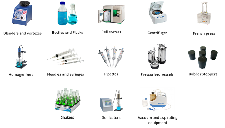
Figure 1: Examples of aerosol producing equipment.
Methods to Minimize the Creation of and Exposure to Aerosols
A combination of the appropriate safety equipment and safe work practices can be used to minimize the creation of and exposure to aerosols.
Control & contain aerosols by following these steps.
- Confine work with biohazards to designated areas.
- Eliminate or reduce aerosol-generating procedures if possible.
- Minimize volumes.
- Use engineered safety equipment to protect personnel from aerosols. Examples include, but are not limited to:
- Certified HEPA-filtered containment equipment, including biological safety cabinets (see Figure 2A), downdraft (animal) cage changing stations, animal bedding dumping stations, aerosol containment tents (“biobubbles”), autopsy/necropsy tables, and vacuums. See biological safety cabinet guidance document for additional information on types and best practices.
- Safety centrifuges with automatic locking mechanisms or solid lids, bioseal rotors, or safety centrifuge cups (see Figure 2B).
- Safety blenders or sonicators.
- Vacuum line traps and filter systems used to protect the vacuum systems.
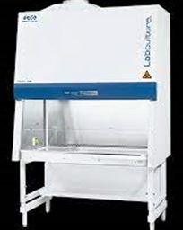
Figure 2: A) HEPA-filtered Class II biological safety cabinet for aerosol containment
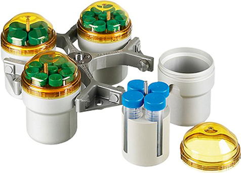
Figure 2:B) Engineered safety cups for aerosol containment
- Adopt safe work practices, including but not limited to the following:
- Biological safety cabinets (see biological safety cabinet guidance document)
- Centrifuging (see centrifuge guidance document)
- Blending, sonicating, grinding, and lyophilizing:
- Operate blender, sonicator, grinder, etc. in a biological safety cabinet if required for the biosafety level of the lab. Otherwise, segregate the procedure to a specific area of the lab, keeping the vessel covered to the extent possible (a towel moistened with disinfectant over the top of blender, grinder, or sonicator is recommended).
- Avoid glass blenders. Otherwise, regularly inspect the bottom of the blender for leakage.
- Open equipment in a biosafety cabinet if required for the biosafety level of the lab. If not, allow aerosols to settle for at least 10 minutes before opening equipment.
- Filter lyophilizer vacuum pump exhaust through HEPA filters or vent into a biological safety cabinet.
- Thoroughly disinfect the area when the process has been completed.
- Autoclave or disinfect all equipment promptly after use.
- Pipetting:
- Mouth pipetting is prohibited; mechanical pipetting devices must be used.
- Pipette all biohazardous materials in a biological safety cabinet if required for the biosafety level of the lab. Otherwise, segregate the procedure to a specific area of the lab, minimizing pipetted volumes to the extent possible.
- Avoid forcibly expelling biohazards from the pipette. Gently drain the pipette with tip against the inner wall of the receiving vessel if possible.
- Place reusable pipettes horizontally in a pan filled with enough liquid disinfectant to completely cover them.
- Thoroughly disinfect the area when the process has been completed.
- Other:
- Use a shielded electric incinerator or hot bead sterilizer to sterilize inoculating loops. Disposable plastic loops and culture needles are good alternatives to open flames.
- Place a pad moistened with disinfectant over the top of rubber stoppers (or similar plug closures) when removing.
- Minimize air bubbles when filling a syringe. Using a passive scoop method or mechanical device, place a pad moistened with disinfectant over the tip of the needle when expelling air.
- If a spill occurs, follow the lab specific spill response plan. Leave the area and allow sufficient time for aerosols to settle. Wearing designated PPE, overlay spill with absorbent material and gently flood spill from outside edge, slowly working toward the center of the spill, to avoid production of aerosols. Avoid forcibly spraying the spill with disinfectant.
- Wear appropriate personal protective equipment according to the lab’s assigned biosafety level or documented risk assessment. If aerosol production cannot be prevented or contained, respiratory protection may be required. Contact EHS Lab Safety Services at ehs_labsafety@utk.edu to determine if the use of a respirator is appropriate.
- Work which may generate infectious aerosols and mitigation strategies must be reported in your Institutional Biosafety Committee (IBC) registration and approved by the IBC.
References
SA0100 – Safety and Environmental Health Program
SA0700 – Safety and Environmental Health Responsibilities
Fleming, Diane O. Hunt, Debra L.. (2006). Biological Safety – Principles and Practices (4th Edition) – 4 Epidemiology of Laboratory-Associated Infections. American Society for Microbiology (ASM).
Gralton J, Tovey E, McLaws ML, Rawlinson WD. The role of particle size in aerosolised pathogen transmission: a review. J Infect. 2011 Jan;62(1):1-13. doi: 10.1016/j.jinf.2010.11.010.
Wooley, Dawn P. Byers, Karen B.. (2017). Biological Safety – Principles and Practices (5th Edition) – 13. Biosafety for Microorganisms Transmitted by the Airborne Route. American Society for Microbiology (ASM).
Disclaimer
The information provided in these guidelines is designed for educational use only and is not a substitute for specific training or experience.
The University of Tennessee Knoxville and the authors of these guidelines assume no liability for any individual’s use of or reliance upon any material contained or referenced herein. The material contained in these guidelines may not be the most current.
This material may be freely distributed for nonprofit educational use. However, if included in publications, written or electronic, attributions must be made to the author. Commercial use of this material is prohibited without express written permission from the author.